Anatomy

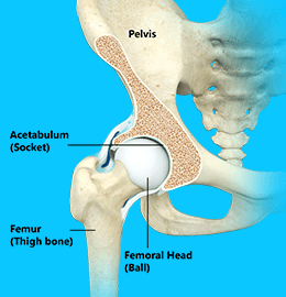
The hip joint is made up of two parts; namely a ball and a socket.
The ball is called the Femoral Head and is the uppermost part of the femur or thigh bone.
The socket is called the Acetabulum and is part of the pelvis.
The ball and socket nature of the hip joint make a very stable construct with a large range of motion.
Surrounding the joint is a joint capsule that allows the joint to be bathed in a special fluid called synovial fluid. This is made by the synovium, a layer of tissue lining the capsule.
Surrounding the capsule are several Ligaments. These are strong, fibrous structures that keep the two parts of the hip joint close together, whilst allowing movement.
Finally, several muscles act across or around the hip joint and are fundamental in making the hip move. The muscles over the front of the hip cause it to flex, and bring your knee up towards your chest. The muscles behind your hip cause your hip to Extend, and and move your leg backwards. Other muscles can rotate your hip in or out, move your leg towards the other one; Adduction, or away from the other leg; Abduction.
Covering the femoral head and lining the acetabulum is Articular Cartilage. Articular means 'of the joint'. It is white and shiny, and you may recognise it if you have prepared a meat joint. The cartilage is smooth and when lubricated with synovial fluid, makes the joint very slippery with very low friction. This means the hip can move many, many times without wearing the cartilage. An average hip will move during walking about 2 million times per year!
When the cartilage is intact, the ligaments and capsule are un-injured and the muscles acting normally, your hip can do wonderful things! When any of the components becomes injured or the cartilage begins to wear out, pain and reduced movement can occur.


